Search This Blog

Sunday, December 13, 2015

How Green Tomatoes Preserve Muscles During Aging

One thing people notice as they age is the loss of muscle mass and strength. This can be accelerated by factors such as lack of exercise, fasting, or an extended stay in a hospital where one is confined to a bed for long periods of time. A recent discovery by a University of Iowa professor of internal medicine and his team may shed some light on the molecular mechanisms of aging and propose two naturally occurring compounds that can help mediate the negative effects of age-related muscle weakness and atrophy.
An old mouse (front) already displaying some physical limitations. Muscle atrophy can be accelerated due to long periods of inactivity. 
(image from Jeff Miller, University of Wisconsin-Madison)

In a paper published in The Journal of Biological Chemistry this past October, senior author Dr. Chris Adams and his colleagues (including Michael Dyle, Steven Bullard, Jason Dierdorff, Daryl Murry, Daniel Fox, Kale Bongers, Vitor Lira, David Meyerholz, Scott Ebert, and John Talley) went through a compound library of 1,309 compounds to look for small molecule candidates that might have potential in mediating muscle atrophy resulting from fasting or spinal cord injury. Life events such as these are major factors leading to muscle atrophy.


Left to right are CM Adams, JJ Talley, and SM Ebert who are co-authors of the paper explaining how they discovered tomatidine and ursolic acid as small molecules that would mediate age-related skeletal muscle weakness. They are now 3 of the 4 key people in a new company called Emmyon, a "biotechnology company that discovers and develops natural and pharmaceutical compounds that improve muscle mass, strength, exercise capacity, and metabolism." (image from Emmyon site)

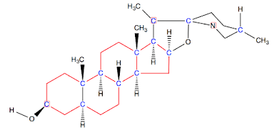
Tomatidine is a naturally occurring compound found in green tomatoes and green apples. Up to 0.5 g of tomatidine can occur per kilogram of fresh green tomato. The compound itself is a steroid as well as an alkaloid.
In this colored image of tomatidine, chiral centers are blue and the steroid part is red. The alkaloid part is on the far right and is classified based on the piperidine carbon and nitrogen skeleton.
Zebra tomatoes are naturally green when ripe and are a good source of tomatidine. 
Tomatidine is a product of hydrolysis from tomatine, a larger molecule that also includes some sugars. This larger compound is thought to play a protective role in tomato plants against fungi, bacteria, viruses, and insects.
Tomatine has been studied for its potential role against fungi, bacteria, viruses, and insects. Can you see which bond breaks in its hydrolysis to get tomatidine?
(image by Edgar181 (Own work) [Public domain], via Wikimedia Commons)

Another compound that the Iowa research team studied for its potential to reduce muscle atrophy is ursolic acid, a compound containing 5 hydrocarbon rings and is known as a triterpenoid. As a triterpenoid, this molecule is made from 6 isoprene units.

 
Ursolic acid, chiral carbons are blue
In a plant, biochemical pathways take 6 isoprene molecules and turn them into a pentacyclic ursolic acid compound.  
(image from http://www.mdpi.com/2072-6651/2/10/2428/htm)
  
 
The peels of Fuji and Granny Smith apples are common sources of ursolic acid.
 (Images by Taken by fir0002 | flagstaffotos.com.auCanon 20D + Sigma 150mm f/2.8 - Own work; by Takeaway - Own work. Licensed under CC BY-SA 4.0)

So what did C. M. Adams and his team discover when adult (6 month) and senior-age (22 month old) mice ingested tomatidine (0.05%) or ursolic acid (0.27%) in their diet? Both had similar effects in that the mice gained skeletal muscle weight by 9-10%, increased the size of type IIb or fast-twitch muscle fibers in the quadriceps, and improved forelimb grip strength by 10-12%

The researchers in another paper suggest that these observations may may due to the inhibitory effect of tomatidine and ursolic acid on ATF4, a transcription factor known to activate several cell stress responses and may be responsible for muscle atrophy. More research is being done.

 
In this fake commercial for Nolan's Cheddar, John Nolan (the animatronics wiz of Where the Wild  Things Are) created a workout for this rat empowered after eating some of the cheese. There is no known publication on the effect of cheese on muscle strength. The video clip is for inspiration (image from https://vimeo.com/12106387).

For More Information:

*Identification and Small Molecule Inhibition of an Activating Transcription Factor 4 (ATF4)-dependent Pathway to Age-related Skeletal Muscle Weakness and Atrophy; SM Ebert, MC Dyle, SA Bullard, JM Dierdorff, DJ Murry, DK Fox, KS Bongers, VA Lira, DK Meyerholz, JJ Talley, and CM Adams; Journal of Biological Chemistry; vol. 290, no. 42, pp 25497-25511.

*Systems-based Discovery of Tomatidine as a Natural Small Molecule Inhibitor of Skeletal Muscle Atrophy; MC Dyle, SM Ebert, DP Cook, SD Kunkel, DK Fox,
KS Bongers, SA Bullard, JM Dierdorff, and CM Adams; Journal of Biological Chemistry; vol. 289, no. 21, pp 14913-14924.

Sunday, November 1, 2015

The Yin Yang of Venoms


In warmer parts of our planet, a snake bite can be traumatic and even fatal without available or cost-effective medical treatment. According to a recent article in Reuters, ~52,000 people have died annually from snake bites in India and Bangladesh, and another ~80,000 people havebeen conservatively estimated in TheAmericas and the Carribean. The highest rate documented is in sub- Saharan Africa where ~100,000 people have died. This statistic focuses on snakes alone and does not includes the insects, frogs, scorpions, lizards, and other venomous creatures people have encountered.

According to the Global Snakebite Initiative, ~2.7 million people in India have been bitten. The lack of educational resources and control programs among poor communities continue to bespeak the need to address this as a global health issue.  
(Image of an Indian cobra from The Global Snakebite Initiative)

Some of the most venomous creatures reside on land and in water:


(Images from https://sciencebasedlife.wordpress.com/2011/04/12/the-most-poisonousvenomous-animals-in-the-world/)


(Images from http://listverse.com/2007/12/16/top-10-animals-you-didnt-know-were-venomous/)

Despite the high incidence of these debilitating and often deadly encounters, there are vastly inadequate or unavailable medical resources. Besides the difficulty of obtaining venom directly from a snake, companies like Sanofi Pasteur which produces the most effective antivenom in Africa typically injects it into sheep and horses. After allowing time for the animals to develop antibodies, researchers extract enough blood and filter out antibodies to the venom and eventually create the antivenoms.


The time and process involved lend to the high cost of treatment – up to $500, an amount most African citizens cannot afford. Because the noted antivenom Fav-Afrique is not profitable for the company, Sanofi Pasteur discontinued manufacturing it last year, and the limited supply of vials is expected to run out some time in June 2016.

While the yin or dark side of venoms highlights may represent their deadliness, it also points to the chemical challenges vaccine manufacturers face in producing these much needed antidotes. Some man-made or synthesized compounds do not last long enough in the body to counteract the venom. In other cases, even venom extracted from the same animal appears differently upon scientific characterization due to the animal’s age, gender, and the environment when it was captured.
  
(image from http://www.ncbi.nlm.nih.gov/pubmed/12955733)
 In the same genus, one can find great variation in venom profiles. These were analyzed by LC/MS (liquid chromatography/mass spectrometry) in the lab of Professor Bryan Fry, aka the Venom Doc, an associate professor at the School of Biological Sciences, University of Queensland.

Then there is the challenge of identifying the molecules or proteins that target specific organs in the victim. These tend to be large (sometimes >65,000 Daltons), compact, and complicated structures tightly held in some cases by disulfide bridges. 
Chlorotoxin is a polypeptide of 36 amino acids with the red alpha helix and blue beta sheet held together by 4 disulfide bridges highlighted in orange. It comes from the death stalker scorpion.  
(image from http://www.mdpi.com/2072-6651/7/4/1079)

The yang side of venoms imparts a different aspect of hope and potential that these compounds may provide medical therapies for debilitating maladies such as diabetes, high blood pressure, chronic pain, and muscular dystrophy. Companies and venom experts collaborate to decipher the complex molecular structures of these peptide molecules and to test their potential for medical application. A few known examples include:
Captopril is used to treat hypertension and congestive heart failure. It comes from the lancehead viper, Bothrops insularis.
 (Top image from https://en.wikipedia.org/wiki/Bothrops_insularis; Left image from https://en.wikipedia.org/wiki/Captopril; Right image from http://www.euvipharm.com/index.php?/en/product/detail/122/captoril)

 
Byetta or exenatide is used in the treatment of diabetes mellitus type 2. It is based on a hormone in the saliva of the gila monster.  
Top left image from http://animals.sandiegozoo.org/animals/gila-monster; top right image from https://commons.wikimedia.org/wiki/File:Byetta_10_mcg.jpg;Bottom image from http://www.polypeptide.com/exenatide-generic-peptides-10.html
Prialt or ziconotide is used to treat chronic pain. 


Ziconotide is very soluble in water. Can you tell why? Look at the number of amine and oxygen-containing functional groups. Also note the S-S bridges that keep this polypeptide together.
 (Top right image from http://www.popsci.com/scitech/article/2005-11/elan-prialt;top left image from http://www.australiangeographic.com.au/news/2014/03/cone-snail-pain-drug-is-non-addictive; bottom image from http://www.mdpi.com/1660-3397/13/8/4967/htm)

Currently, venoms continue to bring hope to those who are personally affected by illnesses with no known treatment. One company, Tonus Therapeutics, was co-founded in 2009 by Jeff Harvey from Buffalo, NY. His grandson J. B. was born with a defective gene leading to Duchenne muscular dystrophy. Without any effective options, Jeff searched online and contacted Frederick Sachs, a professor of physiology and biophysics at SUNY-Buffalo.


Jeff Harvey, co-founder of Tonus Therapeutics

Why Professor Sachs? He had discovered and had been researching the effect of venoms on mechanosensitive channels. The latter are membrane proteins that respond to mechanical stress and can a significant impact on ion channels in the cell membrane.


 (image from https://www.youtube.com/watch?v=__B5isN07AQ)

Here is a nice video description of how mechanosensitive channels work. Note that alpha helices are part of the secondary structure of a protein and play an active role in controlling the channel's size.

Dr. Sach's research involves a venom peptide known as GsMTx4, and it comes from the Chilean rose tarantula. Dr. Sachs and his research team believe this peptide can close the mechanosensitive ion channel that inhibits the flow of Ca ions which can lead to the breakdown of muscle cells.


Chilean rose tarantula, adult male
(image by Viki, http://creativecommons.org/licenses/by-sa/3.0/, via Wikimedia Commons)

(image from http://www.buffalo.edu/home/feature_story/good-venom.html)
Frederick Sachs, a professor of physiology and biophysics, - See more at: http://www.buffalo.edu/home/feature_story/good-venom.html#sthash.ljyGanMq.dpuf

Sunday, October 25, 2015

What's So Funny About Chemistry...

In a super-serious field like chemistry, I sometimes get inspiration from artists who can inject some humor while still seeming sophisticated. Matthew Diffee is one such artist who is a regular cartoonist for The New Yorker and The Texas Monthly. Besides being a talented individual, he is a down to earth Tex-Yorker-Braugh (now in LA) with a convivial curiosity.

image from http://www.matthewdiffee.com/, photo by Scott Gordon Bleicher

Taking inspiration, I thought to try a hand at this as a break from exam week. Feel free to roll your eyes or groan. Once the illness has passed, we'll return to our normal operations.


Sunday, September 20, 2015

A New Way to Figure Out the Age of Old Silk

For museum directors and art experts at places like the Smithsonian Institution and the Metropolitan Museum of Art, art conservation and authentication are vital responsibilities in the valuation of a piece and its relevance in history. This is especially true when the item contains a biological material such as silk. In addition to clothing, silk has been used in art tapestries, flags, and special adornments. Here are a couple of examples:

A fashionable silk coat from the 1700s. 
(images from the Museum of the City of New York - http://www.mcny.org;
http://s481.photobucket.com/user/PiafEdith/media/Habitdeceremonie.jpg.html)


Pair of Cranes on a Branch, from the Freer|Sackler Gallery at the Smithsonian Institution. This Japanese painting was done on silk.
(image from http://www.asia.si.edu/collections/singleObject.cfm?ObjectNumber=F2004.16)
  
The ribbon of this Order of the World War medal is made of silk.
(Image from http://smithsonianscience.si.edu/2011/09/smithsonian-scientists-devise-new-technique-for-dating-silk/)
A common method of dating an object is radioactive carbon-14 dating, where the ratio of carbon-14 / carbon-12 from the object of interest is compared to the ratio found in the atmosphere. Knowing that radioactive decay follows first-order kinetics and that the half-life of carbon-14 is 5730 years enables us to determine the age of the object up to about 60,000 years.

A good primer on the origin of carbon-14 and how it is used to determine the age of something can be seen here:


An example calculation can be found here. 

The problem with using radiocarbon dating is that the sample size for a reasonable analysis should be ~20-50 mg. For a rare and small object, that can be considered too much material to sacrifice to a destructive process. 

To address this concern, Dr. Mehdi Moini (currently an Associate Professor at the Dept. of Forensic Sciences at George Washington University) along with Mary Ballard (Smithsonian Senior Textile Conservator) and Kathryn Klauenberg (Smithsonian Museum Conservation Institute intern) developed a highly sensitive and reliable way to date ancient silk artifacts using capillary electrophoresis mass spectrometry.


Mehdi Moini and Kathryn Klauenberg from The Museum Conservation Institute at the Smithsonian Institution. Between them is the Beckman-Coulter capillary electrophoresis instrumentation interfaced with an LCQ Duo Finnigan mass spectrometer. 
 With this instrumentation, they are able to detect compounds on the attomole scale 
(1 attomole = 10-18 mole)
(image from http://www.si.edu/mci/English/research/technical_studies/ProteomicsResearch.html)

While the concept of using an electric field to separate charged species started in the 60's, automated instrumentation became available in the late 1990's. A nice explanation of the technique is provided by Dr. David Kreller from Georgia Southern University

(image from https://www.youtube.com/watch?v=CXenfe4lMxQ)

What did the research team look for in order to determine the age of a rare silk artifact? It turns out that silk is a long protein molecule composed of many amino acids, and one amino acid in particular - aspartic acid - has the ability to racemize at a fast enough rate to make the analysis reasonable for objects that are less than 2500 years old.
Nonsuperimposable mirror image molecules (enantiomers) of aspartic acid. The chiral carbon and acidic R group are highlighted.

Where do you find aspartic acid in silk?
 Bombyx mori and a silkworm cocoon
(image from http://www.textiletoday.com.bd/oldsite/magazine/574)

It turns out that when you look at an individual fiber of silk, it is composed of 2 kinds of proteins. One is called fibroin which makes up silk's structural center, and the other is sericin which is a gummy substance that coats the outside of silk fibers and enables them to stick together. Aspartic acid is one of three main components of sericin.
(image from http://www.seiren.com/english/products/medical/sericin/)

L-aspartic acid becomes D-aspartic acid over a long time, and by measuring the ratios of D/L isomers in silk samples of known age, Dr. Moini and his colleagues were able to determine the half-life of the racemization to be about 2500 years.

(image from http://pubs.acs.org/doi/abs/10.1021/ac201746u)

Electropherograms of known silk samples showing a greater presence of D-aspartic acid in the oldest samples. 
(image from Anal. Chem., 2011, 83 (19), pp 7579; DOI: 10.1021/ac201746u)

With a 20 minute analysis time using no more than 100 micrograms of silk fibers, the research team developed a method of determining the age of rare silk artifacts without involving significant amounts of material. Because such objects are usually carefully stored in museums and galleries, factors like temperature, humidity, and amount of UV radiation exposure are less problematic.

For further reading:

*Dating Silk By Capillary Electrophoresis Mass Spectrometry; by M. Moini, K. Klauenberg, and M. Ballard; Anal. Chem., 2011, 83 (19), pp 7577–7581;